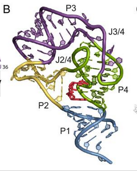
Figure 1. Secondary structure of the c-di-GMP-II riboswitch aptamer domain. P1 is blue, the kink turn (P2) is yellow, P3 is purple, the pseudoknot helix (P4) is green, and the ligand, c-di-GMP is shown in red (1).
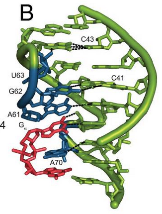
Figure 2. Cyclic-di-GMP bound as part of a triplex. P4 is shown in green, triple helix is shown in blue, ligand is shown in red, and dashed black lines represent hydrogen bonds (1).
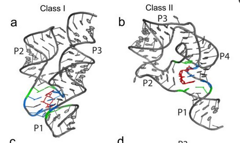
Figure 3. Secondary structures of the Class I and Class II aptamers. The ligand is shown in red, the nucleotides in direct contact with the ligand is shown in blue, and the nucleotides stacked above and below the ligand are shown in green (3).